
What are Skin Lesions?
Skin lesions are a portion of the coating that is different And also starting the skin around it. It may be a lump, a sore, or an area of skin that is not normal. It can also be skin cancer. The skin lesions are of frequent occurrence and may represent a local pathology or manifest a systemic problem. Since the dermis is such a large organ, it is necessary to pay attention to these alterations.
Causes and Tips of Skin Lesions
Frequent Skin Lesions
The skin diseases may have different origins and affect one or more of their structures. We refer to the nails, the hair follicles, and the sebaceous or sweat glands. The specialist indicates to treat them is the dermatologist, who you should visit if you have an alteration.
Acne
It appears at any age but is more common in adolescence. The sebaceous glands fill with detritus, resulting in blackheads, nodules, and blemishes. And also, It is a cosmetic problem, but it can negatively impact your self-esteem.
Prevention consists of keeping the skin very clean by using unique soaps. In addition, there are products design to control the proliferation of the Propionibacterium Acnes bacteria, responsible for the infection of these lesions.
Atopic Dermatitis
It usually makes an appearance in childhood. It is characterize by the presence of plates that. In general, locate symmetrically. You will see them located on the flexor surfaces of the arms and legs, although they also affect the trunk.
It is a kind of eczema that causes redness, itching, and fine scaling. In these cases, the skin lacks certain defenses. Which causes an exceptional sensitivity to irritants. To avoid outbreaks, it is convenient to protect yourself from irritants and use moisturizing and antihistamine products. You should also avoid scratching, as it can worsen the picture.
Vitiligo
This disease is characterize by the appearance of areas of depigmentation and has its origin in the immune system. Which attacks and destroys melanocytes. Lesions can appear anywhere. This depigmentation can even involve the mucous membranes, hair, eyes, and eyelashes.
There is no cure or prevention, but the spots can be alleviated with topical medications and phototherapy. These treatments can include in health insurance and slow down the course of the disease.
Malignant Skin Lesions
Some skin conditions can be malignant or premalignant. Therefore, it is essential that you know and monitors them. In addition, of course, restricting exposure to solar radiation closely relates to its appearance.
Non-melanomatous Lesions
Actinic keratosis, basal cell, and squamous cell carcinoma are found in this group of pathologies. You will see that they are lesions in the form of scaly spots and darker than the rest of the skin. Its origin has to do with solar radiation and the presence of the human papillomavirus or HPV.
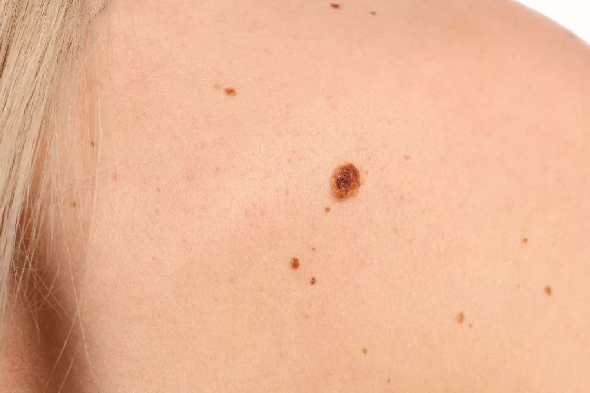
Malignant Melanoma
This skin cancer is common among white people and appears in the areas most exposed to the sun. Such as the face and arms. It is a lesion similar to a mole but with rapid growth, asymmetry, and irregularity in the edges and the pigmentation tone.
The skin lesions involve varying importance. To take care of your health, you need to consult your dermatologist.
Type of Skin Lesions
Primary Morphology
Lip Melanotic Macula
A patch is a large macula. The macules are flat and nonpalpable lesions that usually measure < 10 mm in diameter. They represent a color change and are not uneven from the skin’s surface (neither raised nor depressed). Examples include freckles, flat nevi, tattoos, port-wine stains (hemangiomas), rashes from rickettsia infections, rubella, and measles (papules and plaques may also occur)—some drug allergies.
Skin Lesion (Papule)
The pimples are raised lesions usually measure < 10 mm in diameter that can be felt or felt. Some examples are moles, warts, lichen planus, insect bites, seborrheic and actinic keratoses, some acne lesions, and skin cancers. The term maculopapular often use inappropriately and inaccurately to describe many red-colored skin rashes. Since it is a nonspecific and easily misused term. It should avoid. Abscesses are generally palpable raised lesions of 10mm diameter. Lichen planus (photo) can manifest as a popular rash.
Psoriatic Plaque
The plates typically measure nonpalpable lesions > 10 mm in diameter and are elevated or depressed than the skin surface. The containers can have a flat or domed roof. Basically, lesions of psoriasis and granuloma annular often form plaques. Plaques are palpable raised lesions > 10 mm in diameter. Psoriasis usually manifests as plaques covered with thick, silvery scales of skin.
Secondary Morphology
Trauma-induced injuries, including abrasions caused by the patient’s nails, are typically linear. The configuration is the shape of the individual lesions and the arrangement in groups. The linear lesions arrange straight and said specific contact dermatitis, epidermal nevus linear, and lichen striate.
Skin Lesion (Annular)
The annular lesions form rings with a more apparent central area. Examples include granuloma annular, drug eruptions, dermatophyte infections (e.g., ringworm), and secondary syphilis. Annular lesions appear as rings with a clear central area. And also, granuloma annular is a typical annular skin lesion.
Nummular Dermatitis (Hands)
The nummular lesions are circular or coin-shaped; an example is a nummular eczema. Round erythematous plaques are present on the back of the hands and wrists.
Target Injuries
The target lesions (porthole) appear as rings with a clearer and are classic of the central erythema multiforme. Basically, target lesions (sometimes called iris lesions) manifest as annular lesions with a purplish center and pink halo separated by a pale ring. Such lesions, typical of erythema multiforme, are symmetrically distributed.
Conclusion.
Skin lesions are a joint presentation in primary care, many of which can diagnose based on history and clinical examination. However, the challenge for GPs is distinguishing between benign and malignant lesions so that only those that require urgent review and treatment are referred under the 2-week wait.
And also, the temporary nature of certain types of skin lesions can create confusion in the mind of the inexperienced clinician.
Examination of an animal with a ‘pustular’ disease may characterize clinically by a predominance of papules (preceding the bumps) and crusts (which develop as the pimples dry out and heal). The blemishes may be relatively minor in number and hard to find.
An understanding of the evolutionary process of skin lesions is essential. In addition, a pruritic eruption may transform by chewing and licking into an alopecic. And also, excoriated bleeding patch.
